IMPLANTOLOGY BETWEEN NARROW POSTERIOR TEETH
Dentists
Hospital: Shenzhen University Hospital
Deng, Yongqiang / Director of stomatology department, Shenzhen University Hospital;
Li, Xin/ Post Doctor, HKU Faculty of Dentistry
Li, Xuguang/ graduated from West China School of Stomatology, Sichuan University


Patient information
Patient: Female , 26 years old.
Complaint: Right lower posterior tooth was missing for a year
Medical History: The patient reported no history of hypertension, heart disease , diabetes, myositis ,tuberculosis and other infectious diseases, No history of major trauma or surgery, no history of drug allergy & blood transfusion.
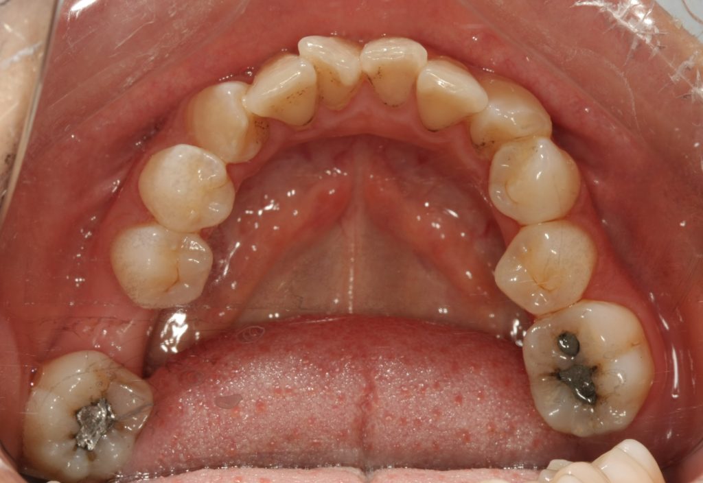
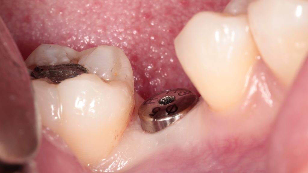
Physical examination: #46 was missing, alveolar ridge shape was blade-like, mucosal color and morphology were normal. the width of the alveolar ridge was about 2mm and the gap in the missing area was acceptable. #47 were inclined to the middle and loose (-)
Auxiliary examination :CBCT show that the alveolar bone in the tooth-missing area was general, The distance between the top of the alveolar ridge and the inferior alveolar neural tube is 19.89mm. No periapical lesions were observed in the adjacent teeth and no abnormal lesions were observed in the alveolar bone
Diagnosis: Tooth-missing
Treatment plan : #46 implant-based restoration.
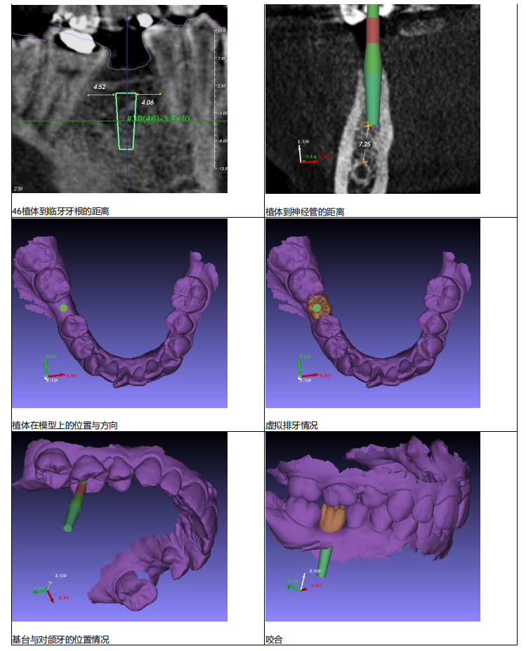
Preoperative CT
-
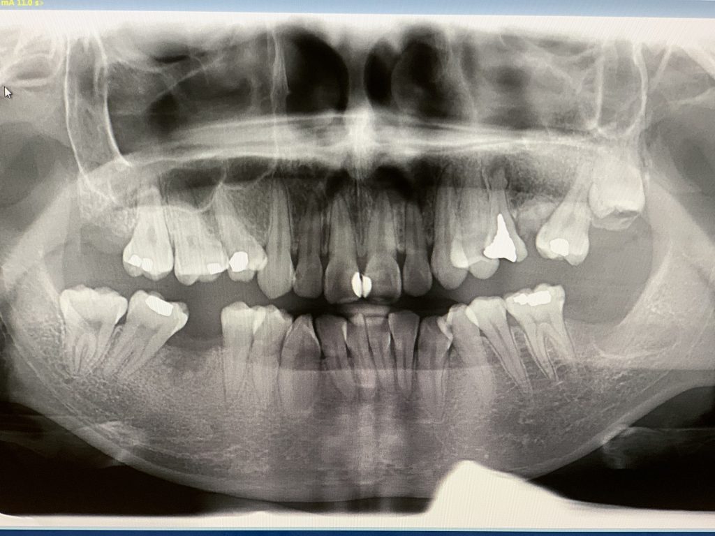
Panoramic X-ray -

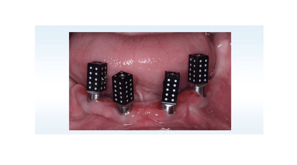
Implant bed region
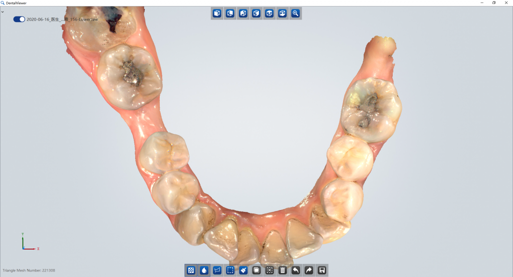
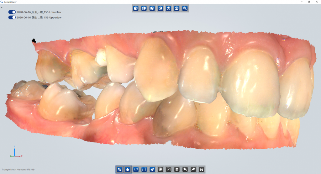
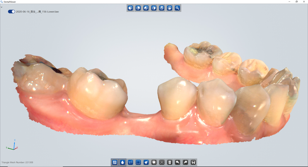
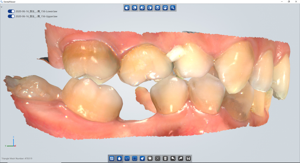
Intraoral Scanning
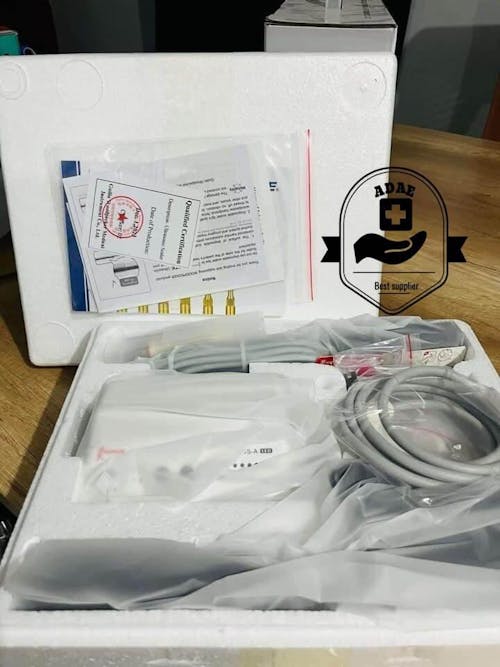
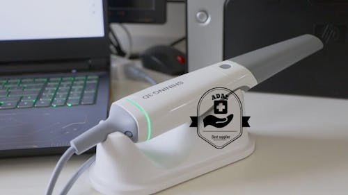
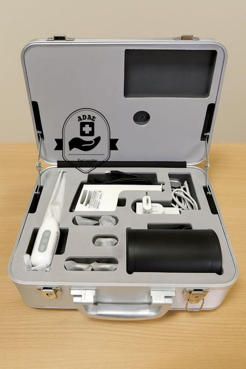
Scan the patient with Aoralscan intraoral scanner. The Aoralscan features true color, easy operation, intelligent scan and sensing motion. Dentists can get the digital impression very easily under excellent infection control.
Operation plan
Tooth position : 30 (FDI #46)
Diameter of implant : 3.70
End diameter of the implant : 2.50
Length of the implant : 11.50
Depth of the preparation : 20
Elongation of drilling : 8.5
Tissue ring cutting :3.70
Sinus lift :0.00
Handle: D2.0 L20.00
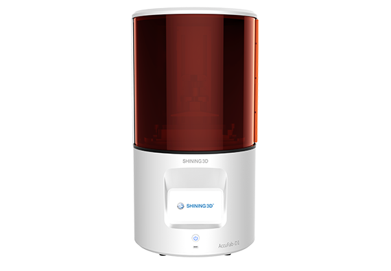
Print Implant Model and Surgical Guide
Print the implant model and surgical guide with AccuFab-D1 dental printer. The AccuFab-D1 supports support automatic creation and one-click printing, offering users very intuitive and user-friendly print experience.
The surgical guide is perfectly fit with the implant model.


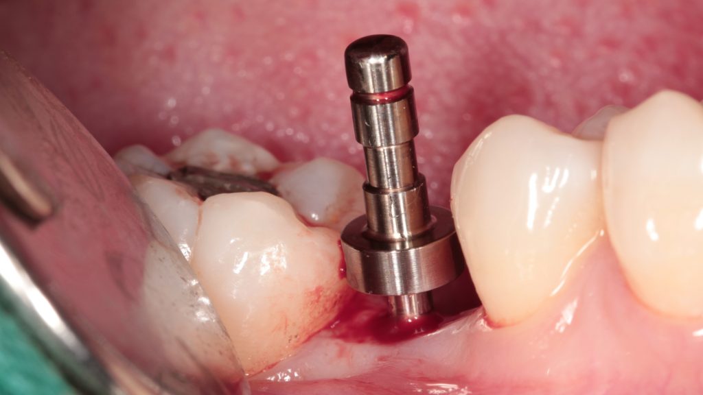
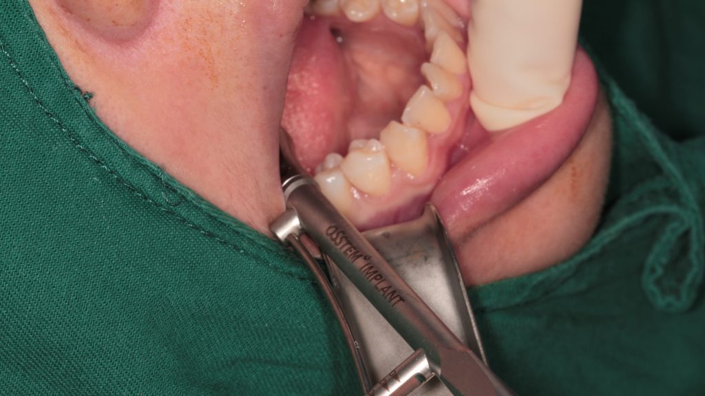
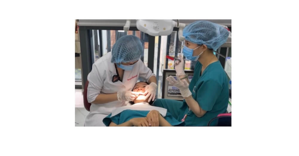
Implant Surgery
Preoperative intraoral view


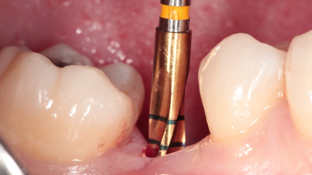
Insert guide in place & prepare implant bed with pilot drill. Check the depth & orientation of the implant bed after drilling.
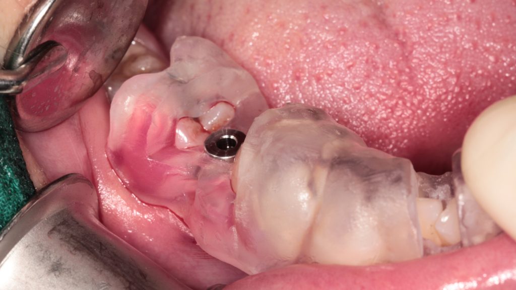
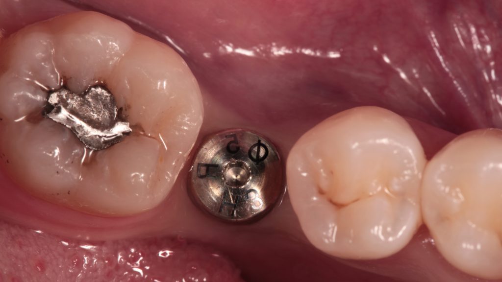
Finish drilling and place implant.
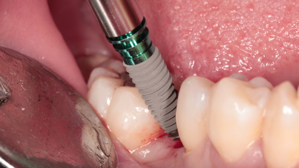
Tighten the implant with torque wrench
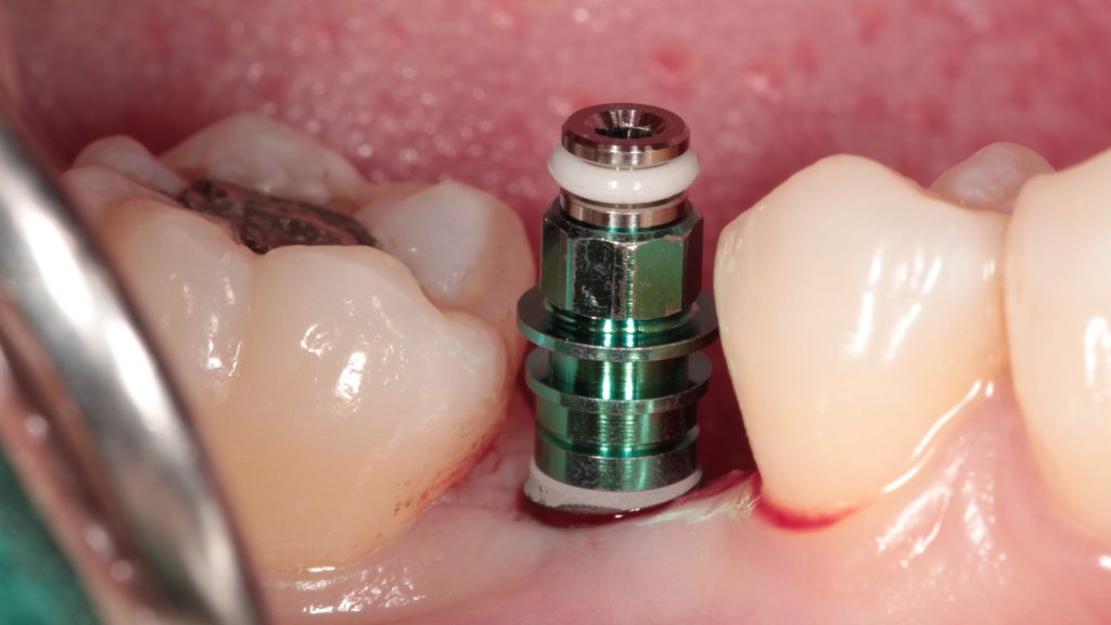
Place healing abutment and complete the surgery.
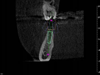
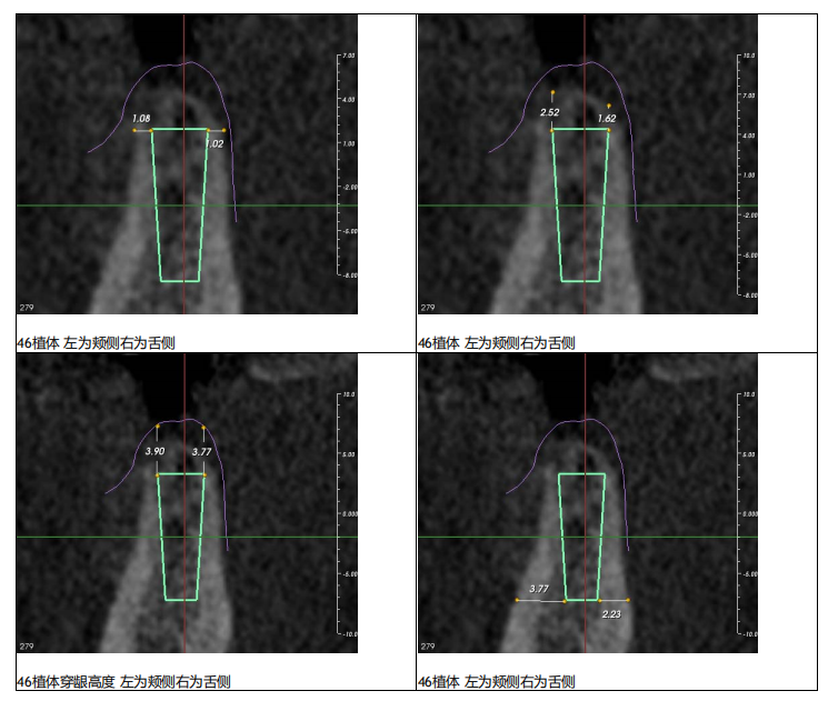
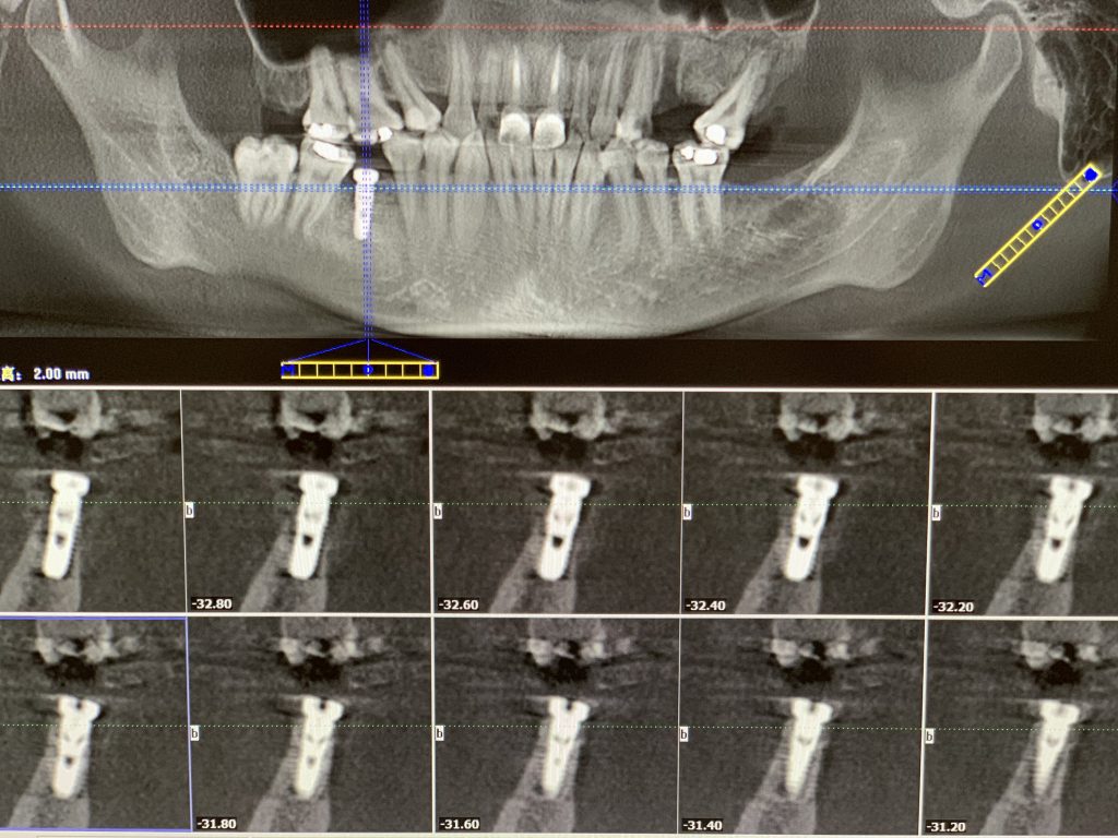
Postoperative CT:
As the buccal & lingual bone absorption plus the superposition of narrow tooth space and other factors, manual implantation is not available. After the use of pilot drill guide for hole preparation, the orientation and depth of the hole preparation can be clearly grasped in the subsequent reaming, which greatly improves the accuracy and safety of the operation


Leave a comment