IMPLANT RESTORATIONS IN TEETH POSTERIOR AREA
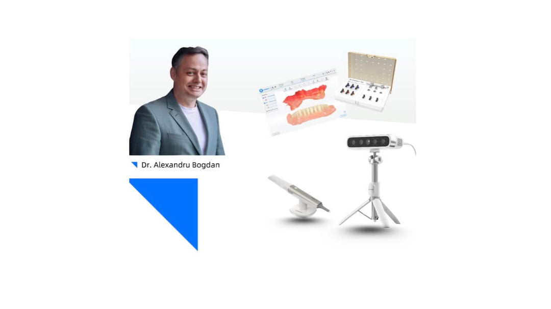
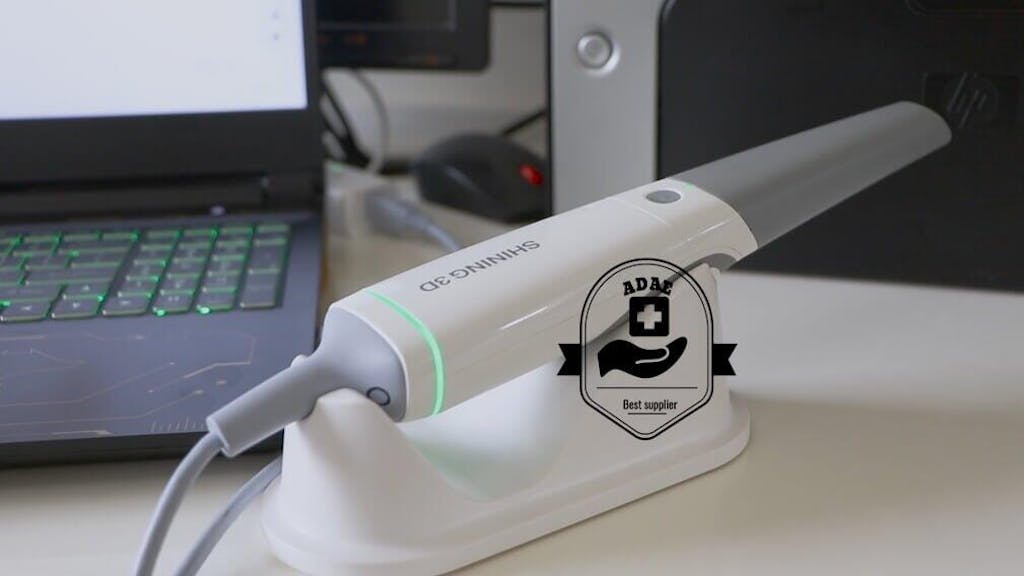
The intraoral scanner has replaced physical impressions in the oral industry in recent years, making it more accurate for dentists and more convenient for patients. The Aoralscan 3, introduced in 2021 by SHINING 3D, has been widely installed worldwide, changing daily dental practice with its elegant device and robust scanning software. Today’s case comes from Dr Abd El-Rahman Khalaf and Dr Kirollos Hany (Fig 1). They are dentists of the Faculty of Dentistry at Assiut University, Egypt. With rich experience, they can provide suitable treatment to patients through digital technology to talk about teeth posterior rehabilitation.

Fig 1: Dr. Abdelrahman Khalaf and Dr. Kirollos Hany are from the Faculty of Dentistry at Assiut University in Egypt
Case Profile
The patient has been experiencing posterior teeth loss in the upper jaw for a prolonged period, resulting in difficulty with chewing. Seeking treatment, he hopes to have stable restorations that will last long-term.
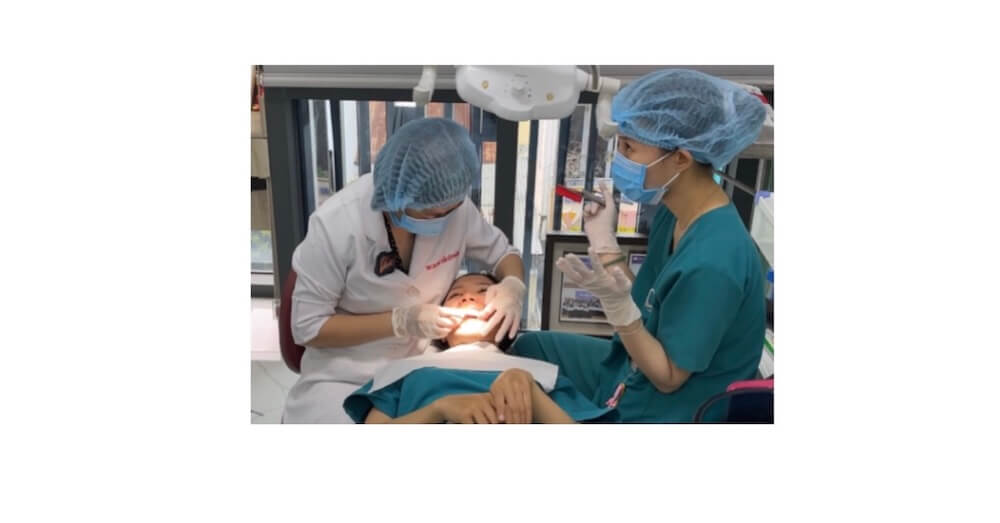
Treatment Process: Implant Surgery
After successful implant surgery and a few months of recovery, the osseointegration between the implant body and the mandible has been established, indicating favourable conditions for proceeding with implant-supported restorations.
Treatment Process: Intraoral Scan
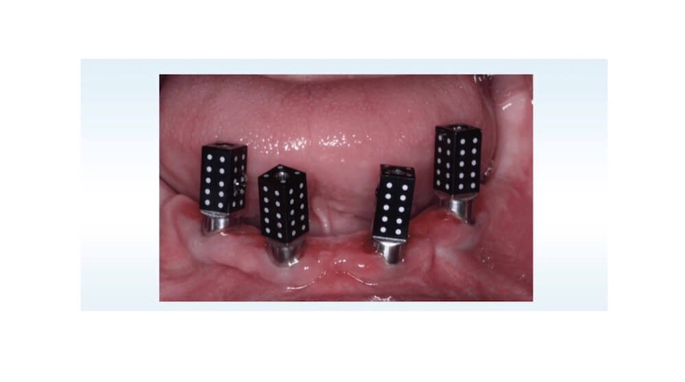
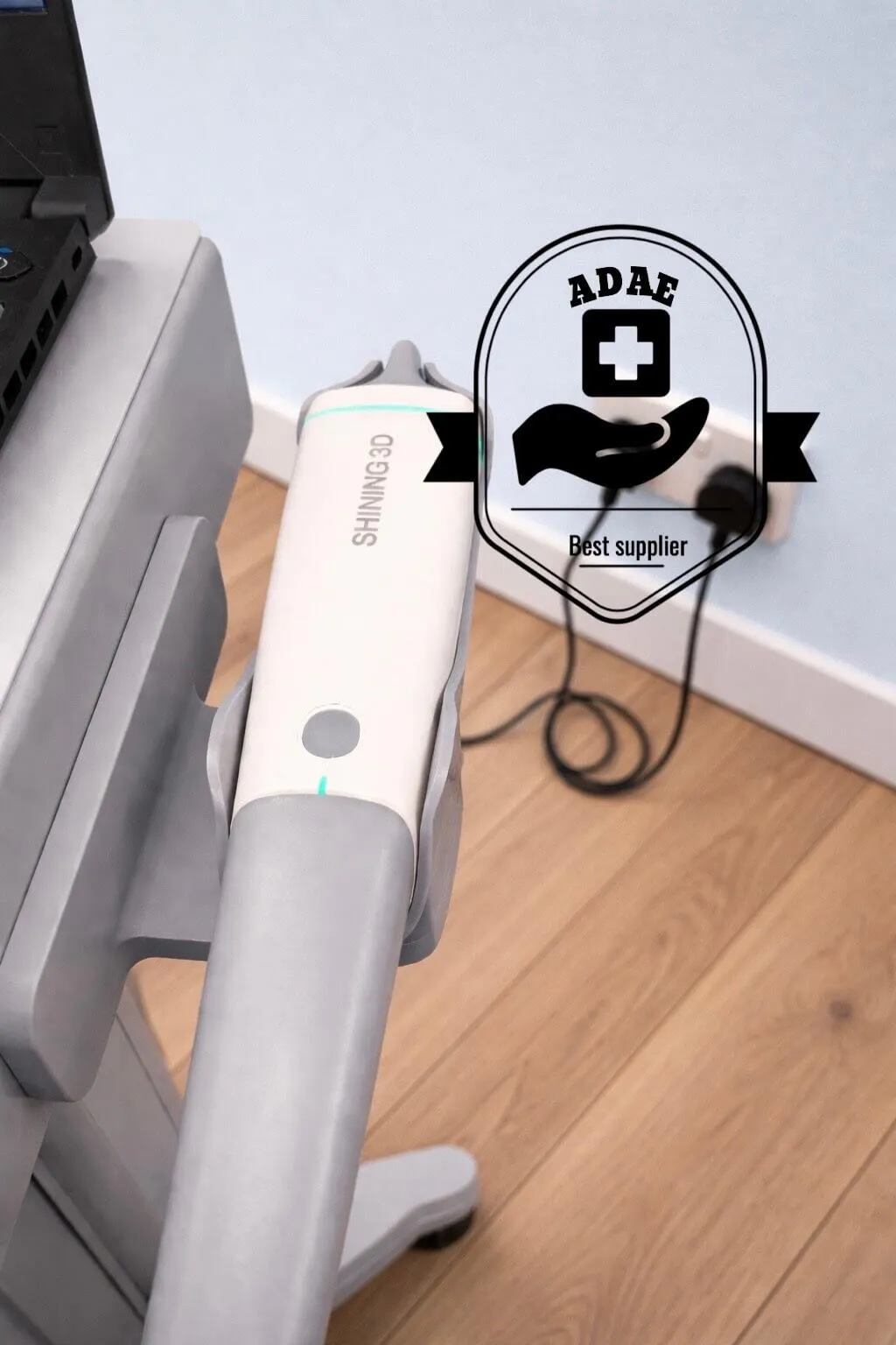
The dentist started by scanning the patient’s intraoral data using Aoralscan 3 without a scan body for the first time (Fig 2). Then, they dug holes to remove the data in the implant position and placed a scan body in the mouth (Fig 3). They scanned again to capture more explicit data of the scan body, then proceeded with the bite scan.
Fig 2, 3, 4, 5: The intraoral scan data captured by Aoralscan 3.
Treatment Process: Design and Printing
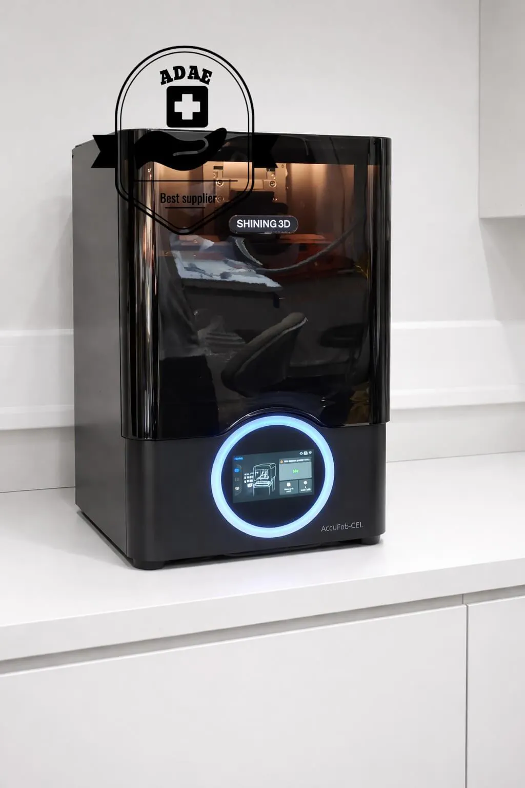
After being scanned, the model was imported into exocad for further design. A single crown and a three-unit bridge were created (Fig 6, Fig 7), and the model was printed using AccuFab-D1s with DM12 resin (Fig 8). The AccuFab-D1s is a highly accurate printer designed for easy use and low maintenance, boasting an incredibly fast printing speed.
Fig 6,7:The restorations were designed in exocad.
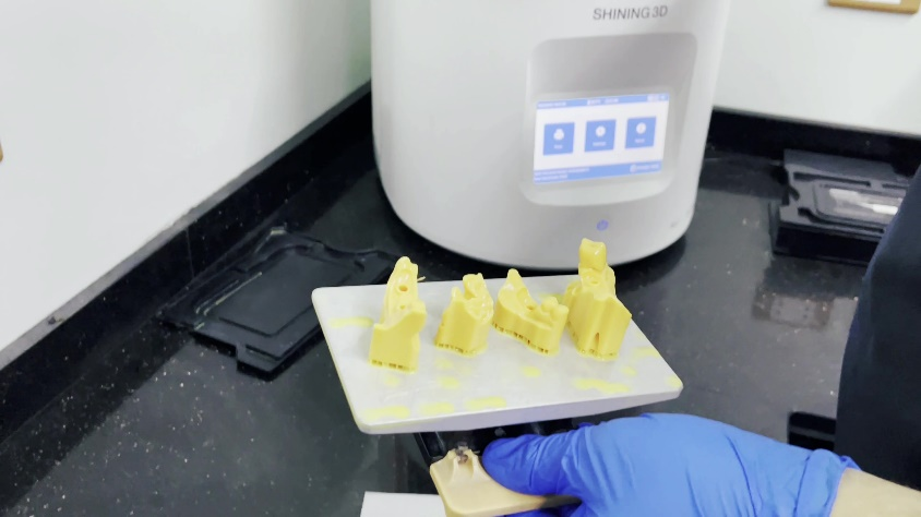
Fig 8: The model was printed by AccuFab-D1s.
Treatment Process: Final Restoration
The final restorations, milled from zirconia, fit well into the model (Fig 9,10). Should any adjustments be necessary, the dentist can make them now before the patient’s arrival.
With no further adjustment about occlusion, shape and adjacency, the restorations were placed in the patient’s mouth, the patient was very satisfied with the treatment result.
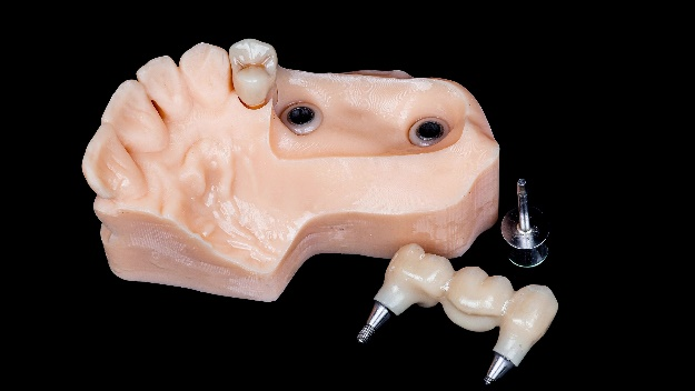
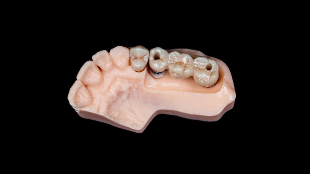
Fig 9, 10: The final restorations in the model.
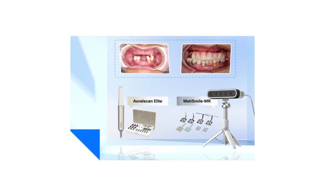
Fig 11,12,13: The result of teeth posterior rehabilitation
Comments from Dr Abd El-Rahman Khalaf and Dr Kirollos Hany
The Aoralscan 3 intraoral scanner used in this case effectively captures precise oral data, ensuring an accurate design of the restorations. This not only increases working efficiency by reducing the need for restoration adjustments during the try-in step but also brings satisfaction to both dentists and patients


Leave a comment